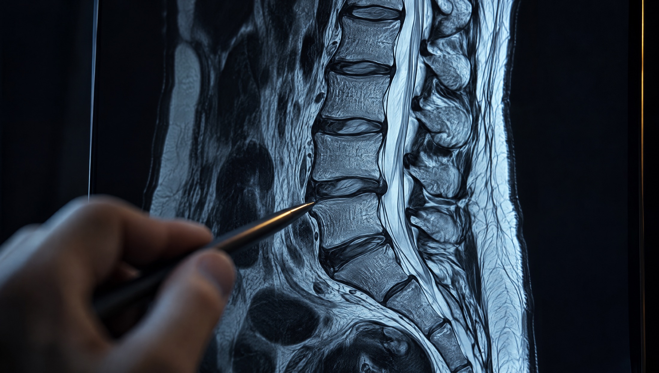
MRI for injury patients.
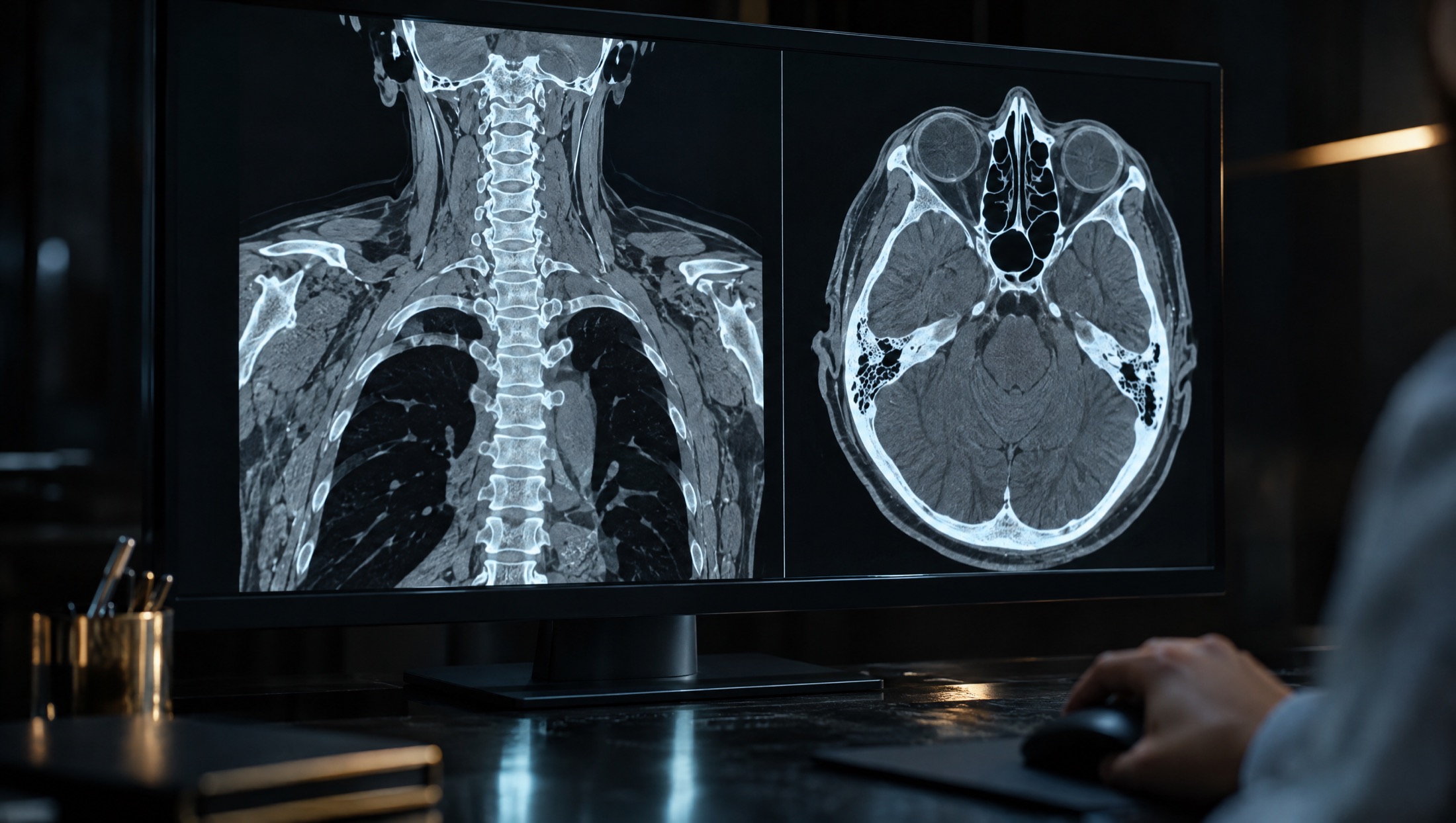
MRI uses a strong magnetic field, not radiation, to make detailed images of soft tissue. It is the study that shows the injuries X-ray and CT usually miss: discs, ligaments, tendons, cartilage, nerves and the brain.
What it can show.
- Herniated, bulging or torn spinal discs and pinched nerves after whiplash or a lifting injury
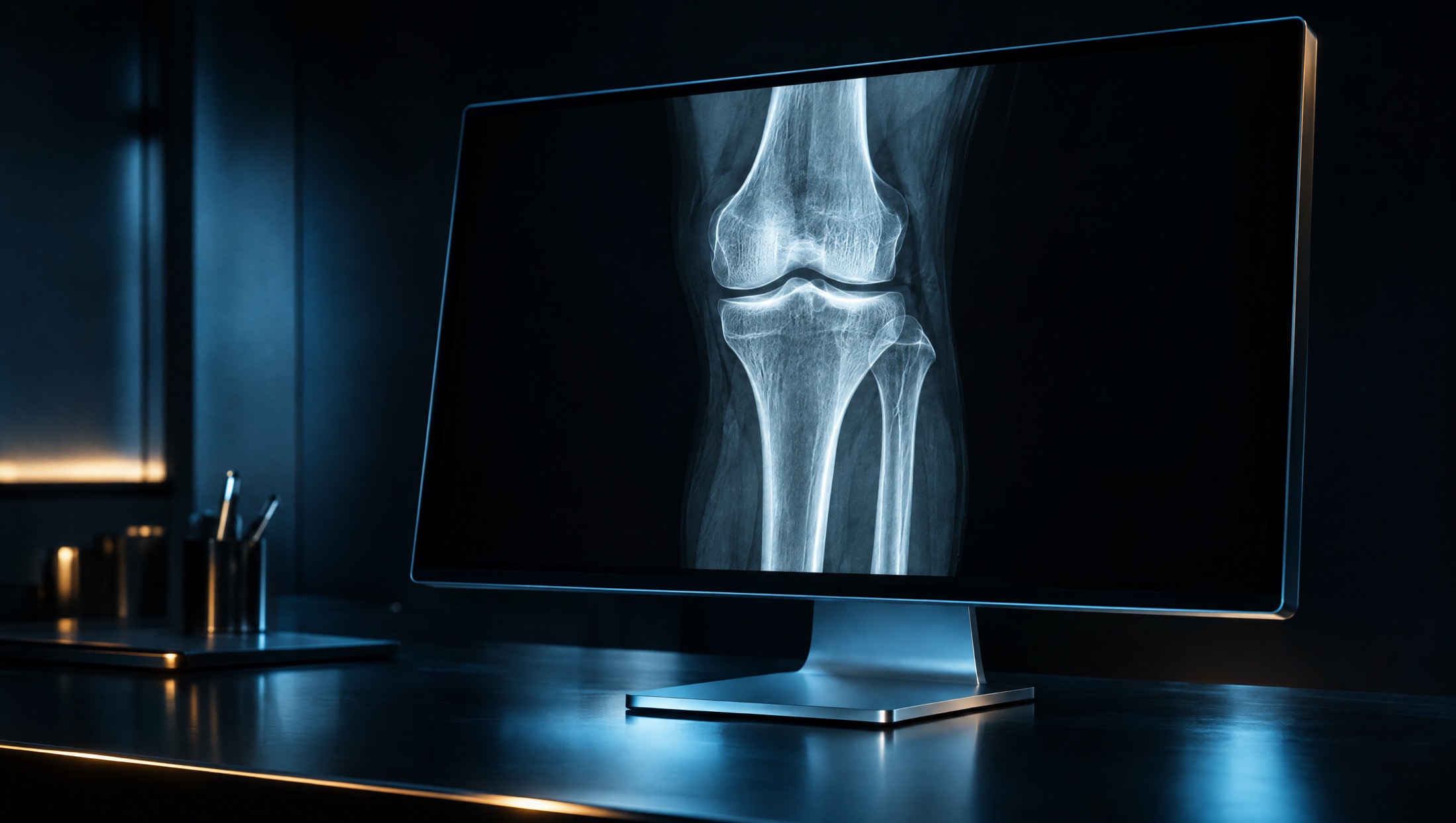
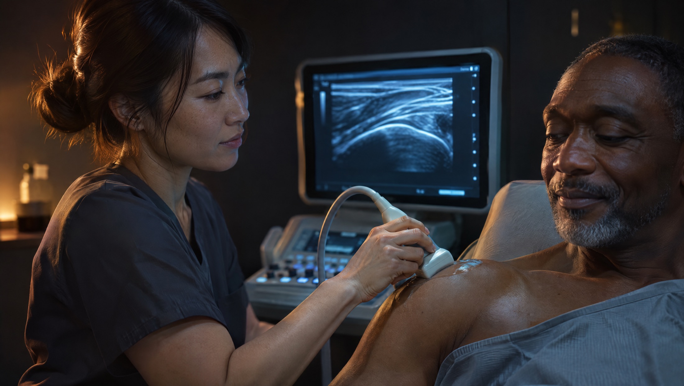
- Torn ligaments, tendons and cartilage in the shoulder, knee, hip, wrist, ankle and elbow, including rotator cuff, ACL and meniscus tears
- Bone bruises and hidden fractures that do not show on X-ray
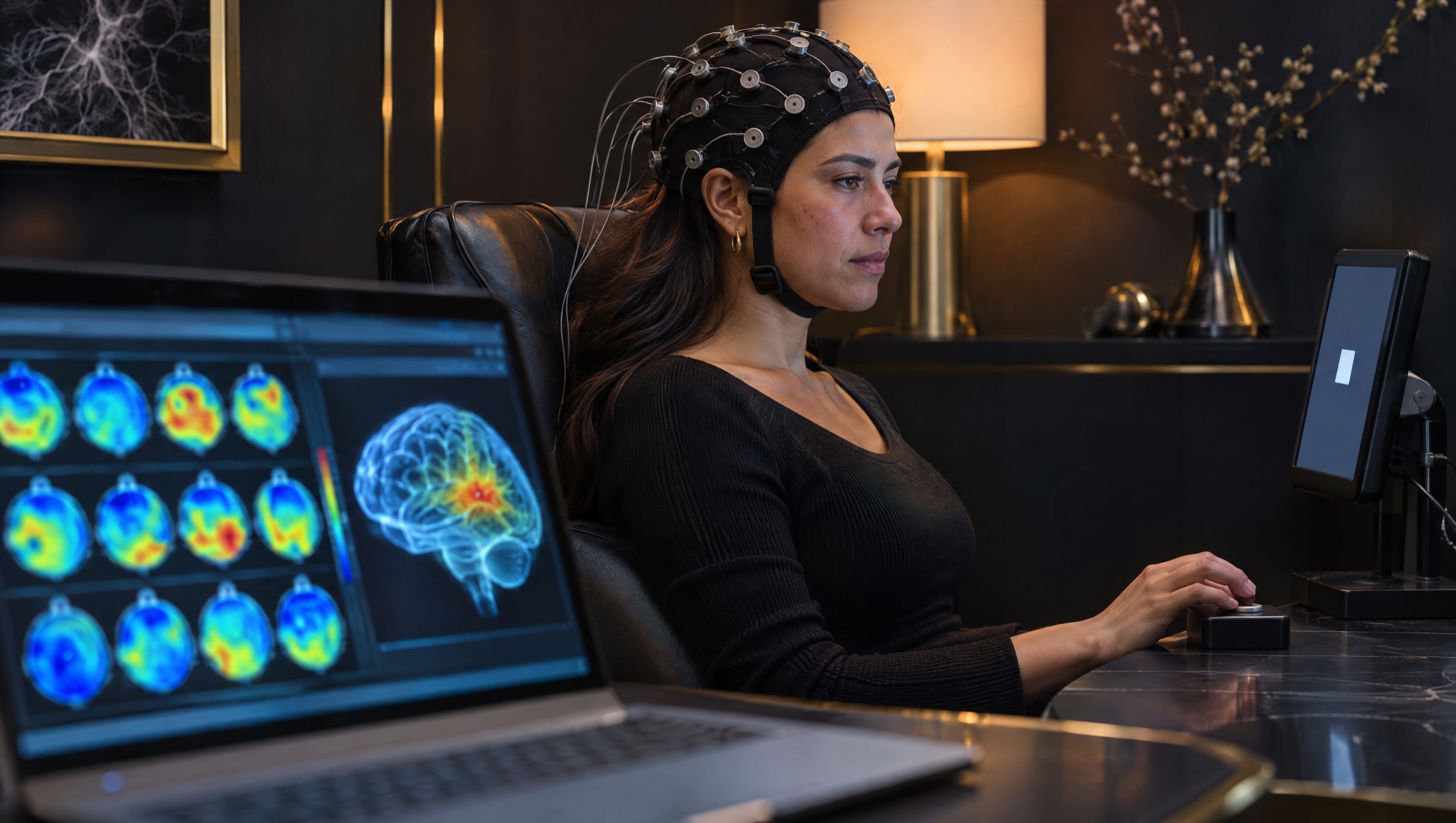
- Bleeding, swelling and other changes in the brain after a head injury
Why it matters to the case
Soft-tissue injuries cause much of the pain after an accident, and MRI is the clearest way to document them.
A detailed radiologist’s report connects the patient’s symptoms to a visible, measurable finding the treating physician can act on.
What the visit is like
Most exams take 20 to 60 minutes, depending on the body area.
You lie still on a padded table while the scanner takes images. It is loud, so we give you earplugs or headphones.
Everything metal comes off before the scan, and we screen every patient for implants first.
Read the full preparation guide